
Physiology of Muscle Contraction
Introduction
Muscle contraction is the physiological process through which muscle fibres generate force. In skeletal muscles, this process allows the body to perform activities such as walking, lifting objects, maintaining posture and moving joints.
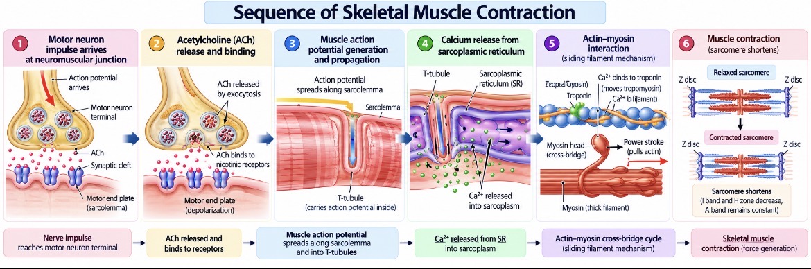
Skeletal muscle contraction begins when a motor neuron sends a signal to a muscle fibre. This electrical signal triggers the release of calcium ions inside the muscle cell. Calcium enables the contractile proteins actin and myosin to interact, producing force.
The mechanism of contraction is explained by the sliding filament theory, which states that thin actin filaments slide past thick myosin filaments, shortening the sarcomere without the filaments themselves becoming shorter.
Real-life example: When you bend your elbow to lift a glass, the biceps brachii contracts and generates force that moves the forearm.
1. Neuromuscular Junction
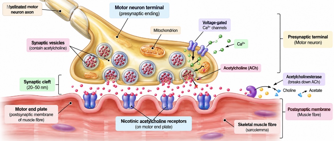
The neuromuscular junction (NMJ) is the specialized connection between the terminal of a motor neuron and a skeletal muscle fibre. It allows a nerve signal to initiate muscle excitation.
The neuromuscular junction consists of three main components.
Motor neuron terminal: The end of the motor neuron contains synaptic vesicles filled with the neurotransmitter acetylcholine (ACh).
Synaptic cleft: This is the small gap between the motor neuron terminal and the muscle fibre membrane.
Motor end plate: This is the specialized region of the muscle fibre membrane, called the sarcolemma, that contains receptors for acetylcholine.
Transmission of the Nerve Signal
When an action potential reaches the motor neuron terminal, voltage-gated calcium channels open, allowing calcium ions to enter the terminal. This triggers the release of acetylcholine into the synaptic cleft.
Acetylcholine diffuses across the cleft and binds to nicotinic acetylcholine receptors on the motor end plate. These receptors open ion channels, producing a change in membrane potential. If the threshold is reached, a muscle action potential is generated and spreads along the sarcolemma.
Acetylcholine is then broken down by the enzyme acetylcholinesterase, which helps terminate the signal at the neuromuscular junction.
2. Excitation–Contraction Coupling
Excitation–contraction coupling is the process by which an electrical signal in a muscle fibre triggers contraction.
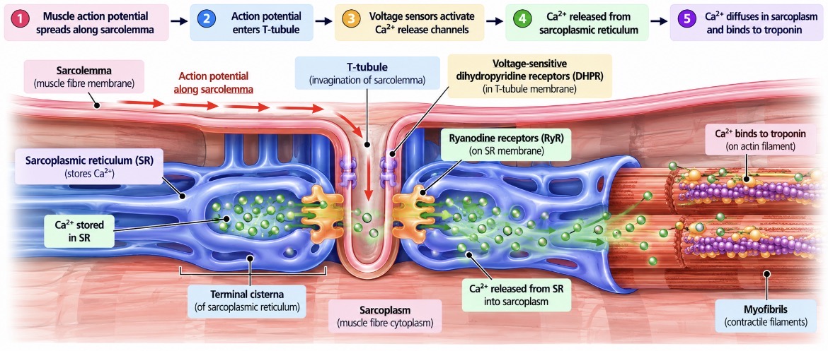
Once the muscle action potential spreads along the sarcolemma, it travels into the muscle fibre through structures called transverse tubules (T-tubules).
The electrical signal activates voltage-sensitive proteins associated with the T-tubules. These trigger the opening of calcium-release channels in the sarcoplasmic reticulum, a specialized internal membrane system that stores calcium ions.
Calcium is released into the sarcoplasm, where it interacts with the regulatory proteins on the thin filaments. This allows actin and myosin to form cross-bridges and generate force.
The main sequence is:
A muscle action potential spreads along the sarcolemma.
The action potential travels into the fibre through T-tubules.
The sarcoplasmic reticulum releases calcium ions.
Calcium binds to troponin on the thin filaments.
Tropomyosin moves away from the myosin-binding sites on actin.
Myosin binds to actin and begins the cross-bridge cycle.
3. Sliding Filament Theory
The sliding filament theory explains how skeletal muscle produces contraction at the sarcomere level.
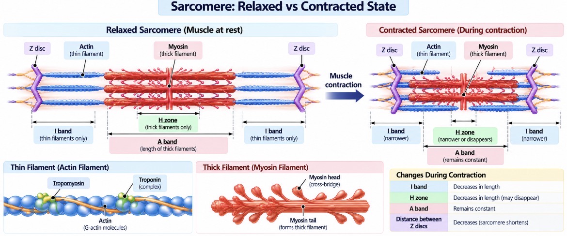
A sarcomere extends from one Z disc to the next. It contains thin filaments, mainly composed of actin, and thick filaments, composed of myosin.
When calcium exposes the binding sites on actin, myosin heads attach to actin and pull the thin filaments towards the centre of the sarcomere. Repeated interactions cause the thin filaments to slide past the thick filaments.
As contraction proceeds, the Z discs move closer together and the sarcomere shortens.
Changes During Contraction
The I band becomes narrower.
The H zone becomes narrower and may disappear.
The A band remains essentially constant in length.
The distance between the Z discs decreases.
The thick and thin filaments do not shorten themselves; their relative positions change as they slide past one another.
4. Role of Calcium in Muscle Contraction
Calcium ions regulate the interaction between actin and myosin.
In a resting skeletal muscle, tropomyosin covers the myosin-binding sites on actin. Troponin is associated with tropomyosin and helps regulate its position.
When calcium is released from the sarcoplasmic reticulum, it binds to troponin C. This changes the shape of the troponin complex and shifts tropomyosin away from the binding sites on actin.
Myosin heads can then bind to actin and generate force.
When calcium concentration in the sarcoplasm falls, calcium dissociates from troponin. Tropomyosin returns to a position that blocks the binding sites, reducing cross-bridge formation and allowing relaxation.
Important: Calcium exposes the binding sites on actin; ATP supplies energy for the cross-bridge cycle and helps myosin detach from actin.
5. Cross-Bridge Cycle
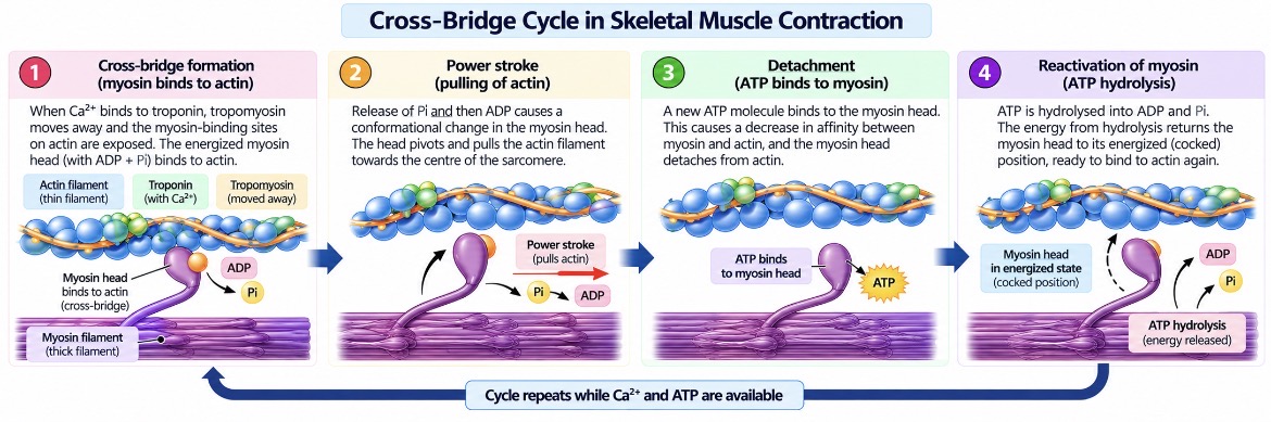
The cross-bridge cycle describes the repeated interactions between myosin heads and actin that produce muscle force.
Step 1: Cross-Bridge Formation
When the binding sites on actin are exposed, an energized myosin head attaches to actin and forms a cross-bridge.
Step 2: Power Stroke
The myosin head changes its position and pulls the actin filament towards the centre of the sarcomere. This movement is called the power stroke.
The release of phosphate and then ADP is associated with the force-producing transition.
Step 3: Detachment
A new ATP molecule binds to the myosin head, causing myosin to detach from actin.
Without ATP, myosin cannot detach normally from actin.
Step 4: Reactivation of Myosin
Myosin hydrolyses ATP into ADP and inorganic phosphate. The released energy returns the myosin head to an energized position, ready to bind again if the actin-binding sites remain exposed.
The cycle repeats while calcium remains available and ATP is supplied.
6. Role of ATP in Muscle Contraction
Adenosine triphosphate (ATP) is essential for normal muscle contraction and relaxation. It supports several processes.
Myosin detachment: ATP binds to the myosin head, allowing it to detach from actin.
Myosin reactivation: ATP hydrolysis provides energy to return the myosin head to its energized state.
Calcium reuptake: ATP powers calcium pumps in the sarcoplasmic reticulum, which transport calcium back into storage and promote relaxation.
Maintenance of cellular conditions: ATP supports ion pumps that help maintain the electrical conditions required for muscle excitability.
Muscle cells regenerate ATP through several energy systems, including phosphocreatine breakdown, glycolysis and aerobic metabolism.
Real-life example: During a sprint, muscles use rapidly available energy sources to regenerate ATP. During prolonged activity, aerobic metabolism contributes substantially to ongoing ATP production.
Important: ATP is required for both contraction and relaxation. When ATP is severely depleted after death, myosin cannot detach normally from actin, contributing to rigor mortis.
7. Muscle Relaxation
Muscle relaxation occurs when the activation of the contractile proteins ends and muscle tension decreases.
When nerve stimulation stops, acetylcholine release ceases and the neurotransmitter already present in the synaptic cleft is broken down by acetylcholinesterase. Muscle action potentials stop being generated.
Calcium-release channels in the sarcoplasmic reticulum close, while ATP-dependent calcium pumps transport calcium from the sarcoplasm back into the sarcoplasmic reticulum.
As calcium concentration falls, calcium dissociates from troponin. Tropomyosin again covers the myosin-binding sites on actin, reducing cross-bridge formation.
The muscle returns towards its resting state through the combined effects of reduced cross-bridge activity and elastic forces within the muscle and associated tissues.
8. Isometric and Isotonic Contractions
Muscles can generate force in different ways depending on whether the muscle changes length and whether movement occurs.
Isometric Contraction
In an isometric contraction, the muscle generates tension without a substantial change in its overall length. The joint angle usually remains relatively constant.
Example: Holding a heavy bag still without moving your arm requires sustained muscle tension.
Isotonic Contraction
In isotonic activity, the muscle changes length while moving a load. Two commonly described forms are:
Concentric contraction: The muscle shortens while generating force, such as when lifting a dumbbell during a biceps curl.
Eccentric contraction: The muscle lengthens while generating force, such as when lowering the dumbbell under control.
Important: These terms describe the mechanical behaviour of the muscle, not different molecular mechanisms. Actin–myosin interactions occur in both shortening and lengthening contractions.
Comparison of Isometric and Isotonic Contractions.
Comparison of Isometric and Isotonic Contractions
Feature | Isometric | Isotonic |
|---|---|---|
Muscle length | Remains approximately constant | Changes |
Muscle tension | Develops | Develops while the muscle changes length |
Visible movement | Usually no joint movement | Movement generally occurs |
Example | Holding a position | Lifting or lowering a dumbbell |
Clinical Significance
Understanding muscle contraction helps explain several physiological and clinical conditions.
Myasthenia gravis: An autoimmune disorder that commonly affects acetylcholine receptors at the neuromuscular junction, causing fluctuating skeletal muscle weakness.
Muscle cramps: Painful, involuntary muscle contractions that can have several causes, including fatigue and altered neuromuscular activity.
Rigor mortis: After death, ATP production ceases. As ATP becomes unavailable, myosin cannot detach normally from actin, causing muscles to become stiff.
These examples demonstrate how normal muscle function depends on effective nerve signalling, calcium regulation and continuous ATP availability.
In summary, skeletal muscle contraction begins with a motor neuron signal and proceeds through muscle excitation, calcium release and repeated actin–myosin interactions. ATP supports the cross-bridge cycle and calcium reuptake, allowing muscles to contract, relax and produce the controlled movements required for everyday activities.