
Appendicular Skeleton: Structure, Bones and Functions
Introduction
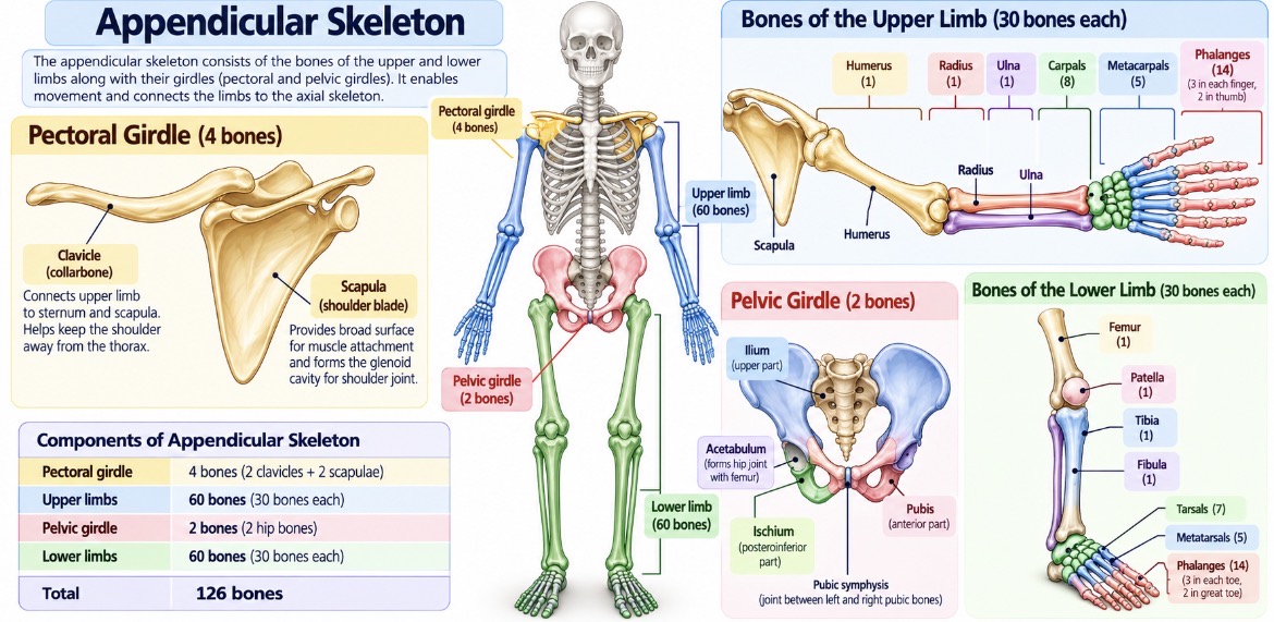
The human skeleton is divided into two major parts: the axial skeleton and the appendicular skeleton. The axial skeleton forms the central axis of the body, while the appendicular skeleton consists of the bones of the upper and lower limbs and the girdles that attach them to the axial skeleton.
In a typical adult, the appendicular skeleton contains 126 bones. These bones allow the body to perform movements such as walking, running, writing, lifting objects and maintaining balance.
The appendicular skeleton is divided into four major components: the pectoral girdle, upper limbs, pelvic girdle and lower limbs.
1. Pectoral Girdle
The pectoral girdle, also called the shoulder girdle, connects the upper limbs to the axial skeleton. It consists of two clavicles and two scapulae, giving a total of four bones.
Its arrangement provides a wide range of movement at the shoulder, allowing the arms to reach, lift, rotate and manipulate objects.
Clavicle
The clavicle, commonly called the collarbone, is an S-shaped bone that extends between the sternum and the scapula. It connects the upper limb to the axial skeleton through the sternoclavicular joint.
The clavicle helps hold the shoulder away from the trunk, transmits forces from the upper limb to the axial skeleton and provides attachment for muscles.
Real-life example: When you raise your arm to reach a shelf, the clavicle moves with the shoulder girdle and helps maintain its position.
Scapula
The scapula, or shoulder blade, is a flat, triangular bone located on the posterior aspect of the upper thorax. It provides attachment for several muscles and contains the glenoid cavity, which articulates with the head of the humerus to form the shoulder joint.
The scapula works with the clavicle and surrounding muscles to stabilize the shoulder and allow a large range of arm movements.
Important: The pectoral girdle is designed for mobility. Its structure permits extensive movement of the upper limb, although this mobility also means that the shoulder is less inherently stable than the hip joint.
2. Bones of the Upper Limb
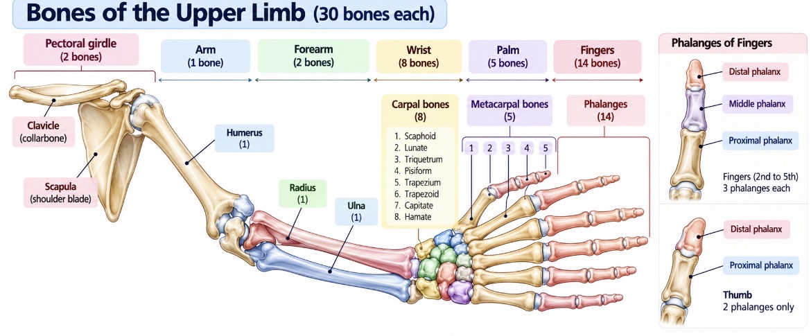
Each upper limb contains 30 bones, excluding the pectoral girdle. Both upper limbs together contain 60 bones.
The bones of each upper limb are organized into the arm, forearm, wrist, palm and fingers.
Humerus
The humerus is the long bone of the arm, extending from the shoulder to the elbow. Its upper end articulates with the glenoid cavity of the scapula, while its lower end participates in the elbow joint.
It provides attachment for muscles involved in shoulder and elbow movements.
Radius and Ulna
The forearm contains two long bones: the radius and ulna.
The radius lies on the thumb side of the forearm in the anatomical position. It plays an important role in wrist movement and rotation of the forearm.
The ulna lies on the little-finger side. Its proximal end forms a major part of the elbow joint and helps provide stability during flexion and extension.
Together, these bones allow movements such as turning the palm upward or downward.
Real-life example: Turning a key or rotating a doorknob involves coordinated movement of the radius and ulna.
Carpals, Metacarpals and Phalanges
The hand contains 27 bones, which provide flexibility, grip and fine control of movements.
Carpal bones: The wrist contains eight carpal bones arranged in two rows. They connect the forearm with the hand and contribute to wrist movement.
Metacarpal bones: Five metacarpals form the framework of the palm. They connect the wrist to the fingers and thumb.
Phalanges: The fingers and thumb contain 14 phalanges. Each finger has three phalanges—proximal, middle and distal—while the thumb has two, proximal and distal.
Real-life example: Holding a pen requires coordinated movement of the wrist, palm and fingers, with the bones providing support for precise muscular movements.
3. Pelvic Girdle
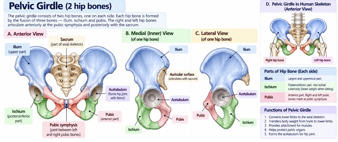
The pelvic girdle connects the lower limbs to the axial skeleton and transfers body weight from the vertebral column to the lower limbs. It consists of two hip bones, one on each side.
Each hip bone develops through the fusion of three bones: the ilium, ischium and pubis.
Ilium
The ilium is the largest and uppermost part of the hip bone. Its broad, expanded region provides attachment for muscles of the trunk and hip.
Ischium
The ischium forms the posteroinferior part of the hip bone. Its ischial tuberosity bears much of the body's weight when sitting.
Pubis
The pubis forms the anterior part of the hip bone. The right and left pubic bones meet at the pubic symphysis, a cartilaginous joint.
The three parts meet at the acetabulum, a cup-shaped cavity that receives the head of the femur to form the hip joint.
The pelvic girdle supports the trunk, helps transfer weight during standing and walking, provides attachment for muscles and contributes to the protection of pelvic organs.
Real-life example: When you stand on one leg, the pelvic girdle and associated muscles help stabilize the pelvis and transfer body weight to the supporting lower limb.
4. Bones of the Lower Limb
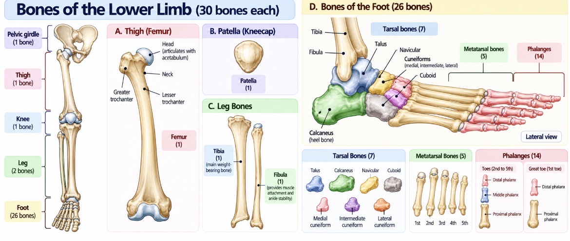
Each lower limb contains 30 bones, excluding the pelvic girdle. Both lower limbs together contain 60 bones.
These bones are adapted to support body weight, maintain balance and produce movements required for standing, walking, running and jumping.
Femur
The femur, or thigh bone, is the longest and strongest bone in the human body. Its head articulates with the acetabulum of the hip bone, while its lower end participates in forming the knee joint.
It supports body weight and provides attachment for powerful muscles of the thigh and hip.
Real-life example: During walking and climbing stairs, the femur transmits forces between the hip and knee and helps support body weight.
Patella
The patella, or kneecap, is a sesamoid bone located within the tendon of the quadriceps femoris muscle at the front of the knee.
It protects the anterior aspect of the knee joint and improves the mechanical leverage of the quadriceps during knee extension.
Tibia and Fibula
The lower leg contains two bones: the tibia and fibula.
The tibia, or shin bone, is the larger, medial bone. It bears most of the weight transmitted through the lower leg and participates in the knee and ankle joints.
The fibula is the thinner, lateral bone. It provides muscle attachment and contributes to the stability of the ankle, but it bears much less body weight than the tibia.
Important: The tibia is the main weight-bearing bone of the lower leg; the fibula primarily contributes to muscle attachment and ankle stability.
Tarsals, Metatarsals and Phalanges
The foot contains 26 bones on each side, organized into the tarsals, metatarsals and phalanges.
Tarsal bones: Seven bones form the ankle and posterior part of the foot. Important examples include the talus, which articulates with the tibia and fibula, and the calcaneus, which forms the heel.
Metatarsal bones: Five long bones form the middle part of the foot and help distribute forces during standing and walking.
Phalanges: Fourteen bones form the toes. The great toe has two phalanges, while each of the other four toes has three.
The bones of the foot, together with ligaments and muscles, help form arches that distribute body weight and absorb forces during movement.
Bone Count of the Appendicular Skeleton
Component | Number of bones |
|---|---|
Pectoral girdle | 4 |
Upper limbs (30 per limb) | 60 |
Pelvic girdle | 2 |
Lower limbs (30 per limb) | 60 |
Total | 126 |
Comparison: Pectoral Girdle and Pelvic Girdle
Feature | Pectoral Girdle | Pelvic Girdle |
|---|---|---|
Bones | Two clavicles and two scapulae | Two hip bones |
Main role | Connects and positions upper limbs | Connects lower limbs and transfers body weight |
Mobility | Allows extensive shoulder movement | Provides strong support and stability |
Important articulation | Glenoid cavity with humerus | Acetabulum with femur |
Everyday example | Reaching overhead | Standing and walking |
Clinical Significance
Understanding the appendicular skeleton is useful for studying common injuries and disorders.
A clavicle fracture can affect shoulder movement. Fractures of the radius or ulna can impair forearm rotation and wrist function. Hip fractures, particularly in older adults, can significantly affect mobility. Injuries involving the tibia, ankle or foot bones may interfere with weight-bearing and walking.
Knowledge of bone landmarks and joint relationships also helps healthcare professionals understand the location of injuries and interpret anatomical findings.
Key Concepts for Examination
The appendicular skeleton contains 126 bones in a typical adult.
The pectoral girdle consists of two clavicles and two scapulae.
Each upper limb contains 30 bones, including the humerus, radius, ulna, carpals, metacarpals and phalanges.
Each hip bone develops through fusion of the ilium, ischium and pubis.
Each lower limb contains 30 bones, including the femur, patella, tibia, fibula, tarsals, metatarsals and phalanges.
The femur is the longest and strongest bone in the human body.
The tibia is the main weight-bearing bone of the lower leg.
The patella protects the knee and improves the leverage of the quadriceps muscle.
In summary, the appendicular skeleton connects the limbs to the body's central framework and enables a wide range of movements. The pectoral girdle supports the mobility of the upper limbs, while the pelvic girdle provides stability and transfers body weight to the lower limbs. Together, these bones are essential for manipulation, locomotion, balance and everyday physical activities.